
|
Forschungsschwerpunkte der Gruppe Rossy (in Englisch)
Our research is at the interface between cell biology and immunology. The questions we want to tackle is how membrane organisation and cell forces regulate T cell activation. To do so, we use mainly advanced fluorescence microscopy and flow cytometry. Past research In the past few years, the lab has focused on understanding how endocytosis and endocytic recycling modulate cell surface expression of the T cell receptor (TCR). TCR is where the T cell-mediated immune response begins and its density at the cell surface, which directly relies on endocytic trafficking, fine tunes the probability and extent of T cell activation. We developed new imaging techniques, based on the use of photoactivatable fluorescent proteins to obtain a dynamic and quantifiable perception of endocytic trafficking in T cells (1).
Video 1: Illustration of the photoactivation approach to quantify the endocytic trafficking of T cell receptor.
In a paper published in 2018, we used this approach to demonstrate that, in contrast to the common belief, TCR is taken up via a clathrin-independent endocytic pathway, which relies on actin polymerisation (2). Because we were able to follow the path of newly internalised receptors with a high temporal resolution, we could show that TCR is incorporated into an endocytic network immediately after endocytosis. This network is defined by flotillins, which are proteins that organise and structure cell membranes. More importantly, our data revealed that flotillins are required for endocytic recycling of TCR and T cell activation. We then pushed further the use of photoactivated fluorescent proteins to generate a comprehensive picture of the entire cycle of endocytic trafficking of TCR in activated T cells. This allowed us to pinpoint the role of flotillins in TCR intracellular trafficking and show that they provide TCR with a “membrane identity” that plays an essential role in sorting TCR for recycling at several stages of its endocytic journey (3). Once in flotillin-positive vesicles, TCR is fast-tracked through a Rab5 and Rab11-positive endocytic axis for a quick return to the immunological synapse. We confirmed the role of Rab5 and Rab11 in this process by using a combination of photoactivation of fluorescent proteins and optogenetic aggregation of Rab5- or Rab11-positive endosomes. Here again, our data question the common belief, which is that fast recycling is mediated by Rab4-positive endosomes. We have conceptualised this model of fast recycling driven by membrane heterogeneity in a recent review (4).
- Ecker M, Redpath GMI, Nicovich PR, Rossy J. Quantitative visualization of endocytic trafficking through photoactivation of fluorescent proteins. Mol Biol Cell (2021) 32:892-902. doi:10.1091/mbc.E20-10-0669.
- Compeer EB, Kraus F, Ecker M, Redpath G, Amiezer M, Rother N, Nicovich PR, Kapoor-Kaushik N, Deng Q, Samson GPB, et al. A mobile endocytic network connects clathrin-independent receptor endocytosis to recycling and promotes T cell activation. Nat Commun (2018) 9:1597. doi:10.1038/s41467-018-04088-w
- Redpath GMI, Ecker M, Kapoor-Kaushik N, Vartoukian H, Carnell M, Kempe D, Biro M, Ariotti N, Rossy J. Flotillins promote T cell receptor sorting through a fast Rab5-Rab11 endocytic recycling axis. Nat Commun (2019) 10:4392. doi:10.1038/s41467-019-12352-w
- Redpath GMI, Betzler VM, Rossatti P, Rossy J. Membrane Heterogeneity Controls Cellular Endocytic Trafficking. Front Cell Dev Biol (2020) 8: doi:10.3389/fcell.2020.00757
- Rossatti P, Ziegler L, Schregle R, Betzler VM, Ecker M, Rossy J. Cdc42 Couples T Cell Receptor Endocytosis to GRAF1-Mediated Tubular Invaginations of the Plasma Membrane. Cells (2019) 8:1388. doi:10.3390/cells8111388
Video 2: Internalisation of TCR in GRAF1 positive tubules (5).
Current projects
The lab is currently organised around three main axes of research.
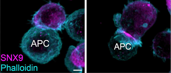
- In the context of a project recently funded by the SNSF, we plan to understand how membrane organisation and endocytic trafficking shape the dendritic cell side of the immunological synapse. We want to pay a special emphasis on the regulation of patterning and density of co-stimulatory and co-inhibitory molecules at the surface of dendritic cells.
- We are developing a cellular system to recapitulate the transition of migration from activation of T cells. This system is based on CHO cells, which we can use a minimalistic antigen presenting cells or as target cells and can be set to represent a T cell activation scenario taking place in a lymph node of in the tumour microenvironment. In combination with a custom analysis, we will use this system to investigate the role of co-stimulatory molecules and chemokines in the probably of T cells to be primed by antigen presenting or kill target cells.
- We have established in the lab two key mechanobiology assays to investigate how immune cells generate forces and how their activity is regulated by cell tension. Current and future projects aim at defining how dendritic cells use force to activate T cells (using traction force microscopy) and how cell tension can promote T cell activation (using a cell stretcher).
About Jérémie Rossy
After completing a master's degree in Biochemistry at the University of Geneva, he focused his research on understanding immune cell signalling. During his PhD at the University of Bern, Jérémie used florescence microscopy to demonstrate that specialised domains are established and maintained in the plasma membrane of leukocytes, and that they control polarisation and migration of these cells. He moved to the University of New South Wales in Sydney in 2009 to develop cutting-edge super-resolution microscopy techniques in order to gain novel insights into early stages of T lymphocyte activation.
Jérémie moved back to Switzerland at the BITG in 2017 and obtained funding from the SNSF (2017, 2021) and the DFG (2020) to pursue his investigations on the role of membrane organisation and mechanobiology in T cell and dendritic cell functions.
A list of Jérémie's publication can be found
here.